Файл:Hepatocellular carcinoma 1.jpg
Hepatocellular_carcinoma_1.jpg (550 × 368 пікселів, розмір файлу: 38 КБ, MIME-тип: image/jpeg)
| Відомості про цей файл містяться на Вікісховищі — централізованому сховищі вільних файлів мультимедіа для використання у проектах Фонду Вікімедіа. |
Опис файлу
| Опис |
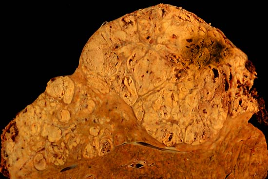
Hepatocellular carcinoma This specimen is from a 50ish woman who presented to the hospital with abdominal pain and ascites. The radiologist recovered what appeared to be whole blood on paracentesis. Cytological exam of the bloody fluid showed no evidence of malignancy. Liver function tests were abnormal, and serologic tests were positive for antibody to hepatitis C. The patient deteriorated rapidly and died within a few days. The autopsy showed this hepatocellular carcinoma occupying much of the volume of a cirrhotic liver. Furthermore, the tumor had invaded the diaphragm and ruptured into the peritoneal cavity, causing the bloody ascites. The photo shows a view of a longitudinal slice taken through the full length of the liver. The photos were shot with a Minolta X-370 with 100 mm bellows lens on Kodak Elite ISO 100 transparency film. The specimen was sliced fresh and fixed in formalin overnight, then briefly immersed in 70% alcohol to retrieve some of the native color and dull the surface reflections. Photograph by Ed Uthman, MD. Public domain. Posted 23 Sep 00 |
| Джерело | http://web2.airmail.net/uthman/specimens/index.html |
| Автор | |
| Ліцензія (Повторне використання цього файлу) |
PD |
Ліцензування
| Ця робота була передана у суспільне надбання її автором, Ed Uthman. Це застосовується по всьому світу. У деяких країнах це не може бути юридично можливо, в такому випадку: Ed Uthman дає кожному право на використання цієї роботи для будь-яких цілей, без будь-яких умов, якщо такі умови не вимагаються за законом.
|
Історія файлу
Клацніть на дату/час, щоб переглянути, як тоді виглядав файл.
| Дата/час | Мініатюра | Розмір об'єкта | Користувач | Коментар | |
|---|---|---|---|---|---|
| поточний | 10:14, 5 червня 2006 | | 550 × 368 (38 КБ) | Patho | {{Information| |Description=Hepatocellular carcinoma This specimen is from a 50ish woman who presented to the hospital with abdominal pain and ascites. The radiologist recovered what appeared to be whole blood on paracentesis. Cytological exam of the blo |
Використання файлу
Така сторінка використовує цей файл:
Глобальне використання файлу
Цей файл використовують такі інші вікі:
- Використання в ar.wikipedia.org
- Використання в ast.wikipedia.org
- Використання в az.wikipedia.org
- Використання в be.wikipedia.org
- Використання в bs.wikipedia.org
- Використання в ca.wikipedia.org
- Використання в cs.wikipedia.org
- Використання в de.wikipedia.org
- Використання в de.wikibooks.org
- Використання в el.wikipedia.org
- Використання в en.wikipedia.org
- Hepatocellular carcinoma
- Alcohol and cancer
- Portal:Medicine/Selected article/50, 2007
- Portal:Medicine/Selected Article Archive (2007)
- Obesity-associated morbidity
- Cirrhosis
- Portal:Viruses/Selected article
- Portal:Viruses/Selected article/10
- User:Daniel Mietchen/Wikidata lists/Items with Disease Ontology IDs
- Використання в eo.wikipedia.org
- Використання в es.wikipedia.org
- Використання в eu.wikipedia.org
- Використання в fa.wikipedia.org
- Використання в fi.wikipedia.org
- Використання в fr.wikipedia.org
- Використання в gl.wikipedia.org
- Використання в he.wikipedia.org
- Використання в hi.wikipedia.org
- Використання в hy.wikipedia.org
- Використання в id.wikipedia.org
- Використання в it.wikipedia.org
- Використання в ja.wikipedia.org
Переглянути сторінку глобального використання цього файлу.